In many dental surgeries, X-ray images that cannot be diagnosed are part of everyday life – even when using digital technology. Bad intraoral X-ray images occur, for example, due to incorrect positioning.
The typical errors with the three imaging media of film, image plate and sensor are often the same:
Distorted images of the tooth because the film or the image plate is bent, a tooth that is not displayed or only partly displayed because the patient moved the film and/or the sensor cannot be correctly positioned (e.g. for flat gums) and/or the incorrect film format was selected.
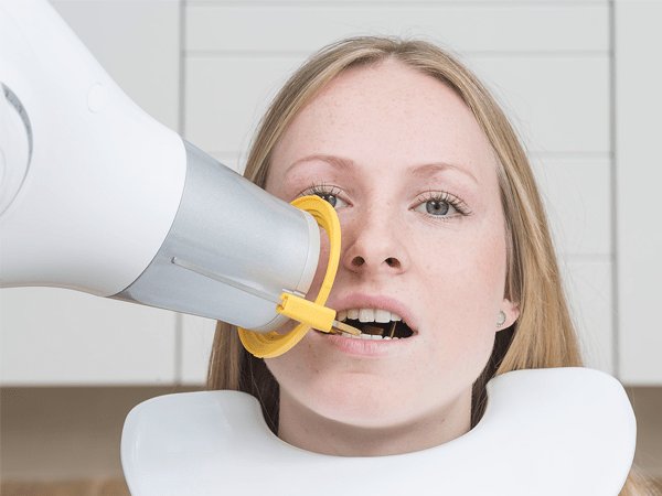
Assistance is provided here by support systems that are positioned using right-angle/parallel techniques. They offer stability and protection against deformation or bending. Using the support systems, precise and reproducible positioning of X-ray film, image plate or sensor can be set, and only little cooperation is required from the patient. As a result, distortion-free images are created without overlapping of the teeth. But not all surgery employees are able to work with the support systems.
Even here there is a potential for error:
The film, image plate or sensor are not fixed in the support so that they can shift in the mouth, the wrong support has been selected for the imaging medium, or the support was incorrectly positioned in the patient's mouth so that he complains of pain. Some surgery teams give up at this point and select the – for them – less burdensome bisecting technique which, however, is also more prone to errors.
Further training in the surgery
In the event of problems with the support systems, surgery trainings for the handling of the right-angle/parallel techniques are possible. These X-ray imaging technique training courses are also offered by Dürr Dental and are carried out directly in the dental surgery. Here, practical tips and tricks are provided in order for the support systems to be used correctly. Because the functioning X-ray imaging technique is purely a matter of practice – as in many areas of daily surgery life.
Of course, the support systems mean additional costs as well as more effort in hygiene since the supports have to be cleaned and disinfected after every patient. But the long-term (also financial) advantages clearly prevail. If properly used, the supports prevent damage to the imaging media.
In other words:
Less scratches on the plate, the film or the sensor, hence longer durability. Because replacing imaging media is significantly more expensive in the long run than acquiring a support system.
An additional use of the support systems is in dosage reduction:
Thanks to the aiming rings with radiation field restriction, intraoral images for children can also be carried out with the recommended radiation field restriction.
Supports assist the surgery team in correct positioning during intraoral X-raying.

