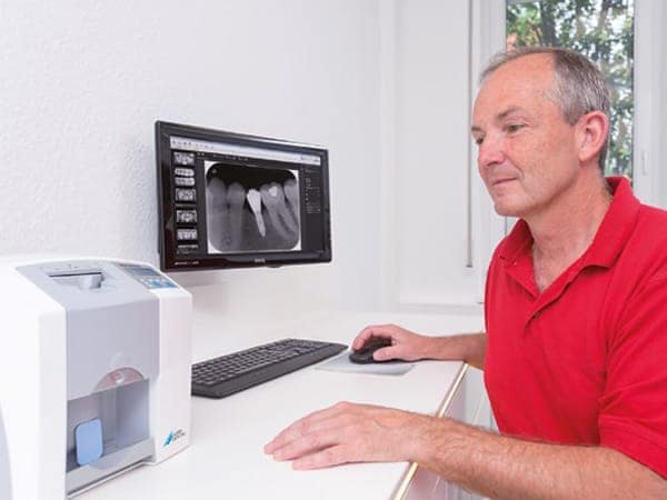
D’life: For you, what are the most important steps in a safe diagnosis of caries and what is the role played by imaging systems?
Every diagnosis of caries begins with a physical examination. Even when presented with indications of caries, for example proximal caries, I still need to use imaging equipment, as only an x-ray will provide final certainty. This is not just my opinion, but a general consensus. Bite wing imaging is not regarded as a proof.
What are the critical points for a reliable caries diagnosis? Are there any circumstances which can impede a diagnosis?
Tooth discolouration for instance. Such anomalies in the dental enamel sometimes make it difficult to differentiate between harmless discolouration and caries. A further challenge is presented by tilting and rotations which even manifest themselves in non-orthodontic cases. Such constellations can result in superimposition of the approximal spaces, which hampers a correct diagnosis. Dental prostheses also pose a considerable problem The sub-gingival ending of bridges or crown edges also often prevent a clear diagnosis. Individual patient characteristics can also play a role. Some patients have an extremely strong pharyngeal reflex, which is often triggered when the dentist trying to position their tongue with a mirror, so as to take a picture of the rear mouth area or the lower jaw. Patients with a high mouth base suffer a similar situation. If this is the case, the bracket required for the parallel technique of bite wing imaging (holding the film) presses into the base of the patient's mouth in such way that prevents them from biting.
You have recently made the change from sensor technology to image plates in your practice. Which advantages do you expect?
Patient feedback indicates that this was a very good decision. The image plate is narrower and more flexible than the sensor, and produces a much more comfortable feeling. Patients also no longer have a cable sticking out of their mouth, which they also disliked. The change with the image plate is easier for the staff to accept if you first change from analogue to digital. This means that the personnel are used to handling film formats and are not forced to start from scratch. This also brings obvious advantages associated with digital x-rays. The recordings can be processed, printed, duplicated or disseminated. For example, an oral surgeon may request a copy. Not to be underestimated is that the practice no longer needs to dispose of the development and fixing fluid. This also deals with a range of environmental problems. The short development time associated with digital x-rays is also a clear advantage. This is important for example, if an x-ray should be taken during an operation to ensure a successful outcome. During a refresher course on diagnostic imaging, the tutor indicated that between 40 and 50 per cent of dentists still use analogue x-rays. I made the changeover to digital technology over 15 years ago, and today would not do without it. For me, this even outweighs the price of the new purchase. Nevertheless, I can understand why many colleagues feel as they do. Viewed in the context of the increasing digitalisation of the dental practice, this measure makes complete sense.
I am fully convinced by the picture quality resulting from the use of » image plates. The difference of the quality which I produced 15 years ago is considerable. The quality of the x-ray picture produced in difficult situations enables me to recognise even the finest structures, something which even helps with a reliable diagnosis of caries. The highly sensitive nature of the image film means that this technology requires lower levels of radiation. A further considerable advantage in comparison with the sensor is the various format sizes which the image film produces. This provides access to even the most inaccessible areas in a patient's mouth. The new technology also brings considerable advantages with children, generating good pictures of very small mouths. A picture taken with a sensor was able to cover a maximum of two teeth. An image plate is able to record three. If subject to proper maintenance, the image film can be reused many times over a long period.
Many thanks for the interview.

