
High-quality X-rays are the basis for reliable dental diagnoses. Two imaging techniques are generally used in intraoral radiography, which require different knowledge and procedures for positioning the patient.
What’s important: The aim of intraoral imaging with imaging plates is to create a complete, detailed and high-resolution image of the affected tooth, including the two neighbouring teeth. The focus is on crowns and roots including the root tips, the periapical region and the periodontium.
Parallel technology X-ray
The assistant uses a holder to align the image plane parallel to the tooth axis. The central beam runs parallel to the guide rod on the holder system. The ring outlines the irradiated area and ensures that the tube points to the image plane at the correct angle. The bite plate fixes the imaging plate securely in the patient's mouth.
- The holder system must be assembled correctly in order to function faultlessly
- If the bite plate is not sufficiently fixed by closing the patient's mouth, in many cases the root tips are not completely imaged
- If certain anatomical conditions are present (e.g. high floor of the mouth or pointed roof of the mouth), the patient cannot close the mouth completely
Tips from practice:
- Dürr Dental provides various videos and educational posters on the website that demonstrate the positioning of the patient and the use of the holder system VistaPosition PSP. You also have the opportunity to register for a team training.
- You can find our English-language webinar here
- If the patient is unable to close the mouth completely when taking pictures in the mandible, in many cases the downward orientation of the bar makes it easier to close the mouth
- Further tips on positioning depend on the type of recording. These recommendations will be dealt with together with the respective topic in the upcoming newsletters
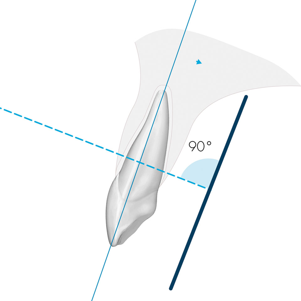
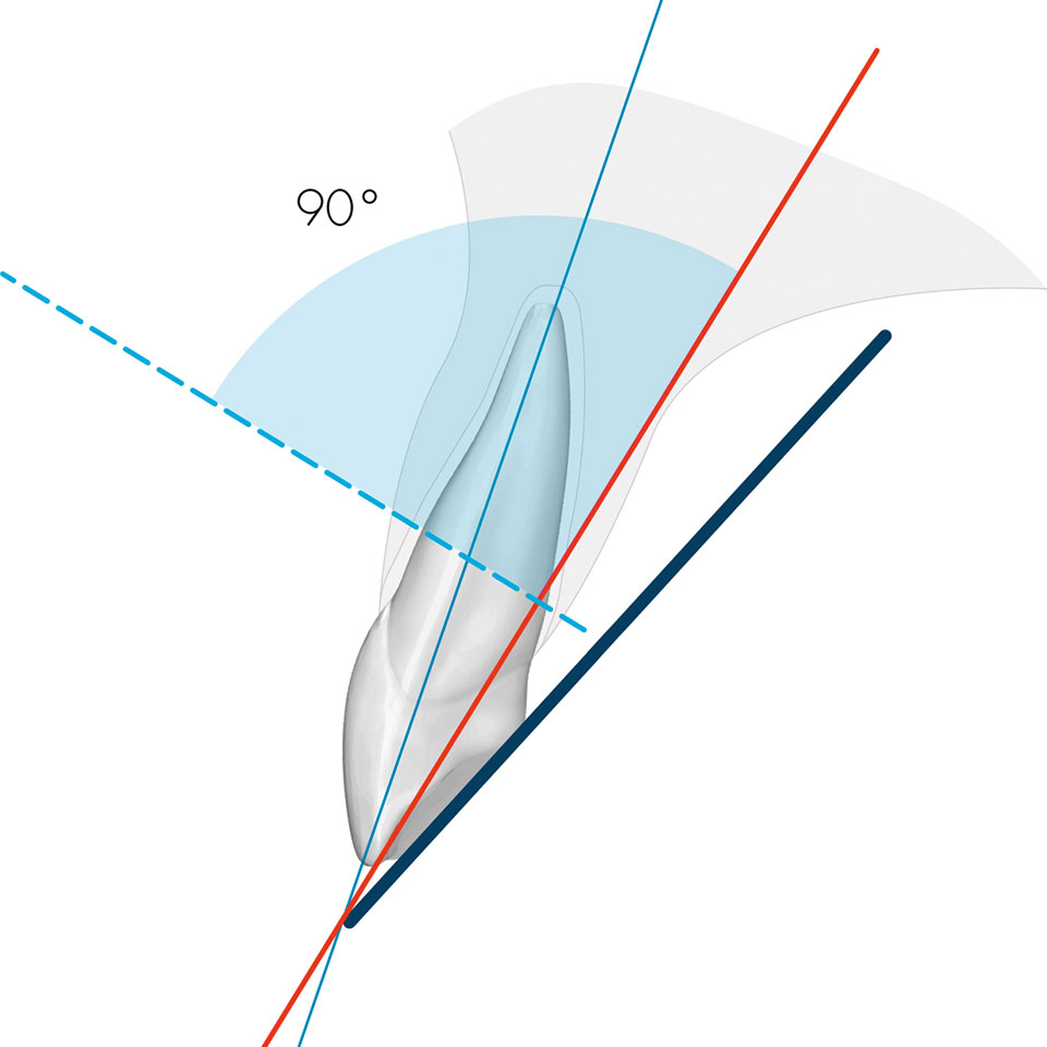
Half-angle X-ray technique
The assistant aligns the occlusal plane of the patient's jaw parallel to the floor. The beam head is aligned according to the degree specification for the region to be imaged. When the image is taken, the central ray strikes the imaginary plane of the ‘angle bisector’ between the tooth axis and the image plane at an angle of 90 degrees. The patient fixes the imaging plate in the mouth with a finger.
- Positioning the patient without a support system requires experience, spatial awareness and anatomical knowledge
- As the patient holds the foil in his or her mouth, it can easily slip and, as a result, important parts are not shown on the image
- Finding the right angle for aligning the tube is very complex. If this is not successful, distortions or white spots will appear on the image if they have not been correctly hit by the X-ray beam
Both recording techniques have their justification. However, the parallel technique with holder is less complicated. Experience has shown that inexperienced dental assistants in particular need more time to familiarise themselves with the half-angle technique. Advantages of parallel technology:
- The parallel technique is easy to learn and requires less experience
- The entire active surface of the image plate is reliably exposed
- The imaging plates can be used longer because the ergonomic design of the bite plate in the holder ensures that they are not bent or crushed
- The image section can be reproduced with regular repetitions of the images