“The case went down as follows”, she tells us. “In 2011 I made an X-ray image of a patient’s tooth 15, which showed a quite resected root. But three years later the patient was again complaining of pain in Regio 15. I prepared another image, this time with the VistaIntra from Dürr Dental, and lo and behold – now a second palatal root was visible!”
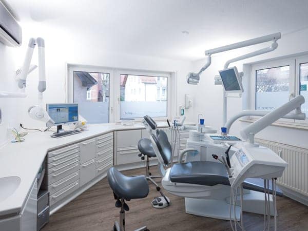
The VistaIntra is one of four digital devices from Dürr Dental that » MUDr. Heda Dengel uses in her surgery to provide her patients with yet better diagnoses. She is following a steady and ongoing trend – comprehensive digitalisation of dental surgeries. In the development of devices, machines and software, as well as in the arrangement of the digital process chain, the market always makes new developments available which, depending on need, can be put together as modules or even as fully digitised surgeries. Surgeries that have grown accustomed to the advantages of digital storage can no longer imagine their workday without digital X-ray and camera technology. High quality digital images are instantly filed in digital storage and made directly available in electronic record cards. The scanner makes manual filing methods superfluous. MUDr. Dengel has long been convinced that digitalisation even for, as she calls it, a “small-town surgery”, is worth it.
More time for patients
She set up her own surgery four years ago in Remseck, near Stuttgart, and has since then been enthusiastically testing the performance of modern surgery software and intelligent imaging systems. The goal is to use digitalisation to optimise work processes and quality management, to reduce costs, become more efficient in administrative work and to gain valuable time – time that will be to the patients’ benefit. “Patient needs stand front and centre for me”, says MUDr. Dengel. “And for my patients digitalisation, above all, means more comfort and service. It is, for example, a huge benefit when during advising and therapy planning I can show patients everything on the monitor”.
She now relies entirely on imaging systems such as the VistaScan Perio. The image-plate scanner enables timesaving digitalisation of image plates. Especially when used in conjunction with VistaIntra, the X-ray tube assembly for intraoral imaging, the image-plate system delivers remarkable image results. “I previously used an older X-raying device in combination with the image-plate scanner. Since working with the VistaIntra I can identify considerably more in the images”, says the dentist.
Documentation and diagnosis
Digitisation also facilitates documentation and diagnosis. A device that MUDr. Dengel regularly uses is the Vista-Cam iX. Thanks to its innovative interchangeable head, the intraoral camera can be used not only for intraoral images, but also for 120-fold enlargement, for diagnosis of fissures and approximal caries or for light hardening. “For one I use the camera in order to show my patients everything on the monitor and to explain the treatments to be applied. For another the VistaCam iX is very helpful for documentation, such as for changes to mucosa. The camera delivers quick images in top quality”, according to Dengel. The proof-interchangeable head is especially useful. “Some teeth show dark fissures” she explains. “Mirrors and probes in and of themselves cannot determine beyond a doubt whether or not there are caries”. She can now spare her patients the previously necessary test drilling. “Checking with VistaCam iX is convincing enough”, she explains.
Precise panorama images
The VistaPano S panorama device was available for testing to MUDr. Dengel before it came to market. “That was unusual because I could exert direct influence with the experience I had gathered in my everyday work” she said. “Even the developer paid us a visit and coordinated with us”.
The product has won her over. “VistaPano S can capture several layers in one cycle, whereas other latest-generation panorama devices require manual adjustment of the sharpest image areas”, says Dengel. From a technical standpoint, the VistaPano S splits every layer into fragments. The most sharply focussed fragments are automatically selected and assembled to create the panorama image. “VistaPano images achieve the best sharpness possible in all image areas because they include individual anatomical factors, as well as momentary patient position, so that features such as roots in the lower- and upperanterior teeth area are sharply depicted”, she explains. “The images are very precise and save me time because I only need to produce one image and no clarifying mouth film”.
For patients, digital systems also provide relief, since radiation doses can be held low. “My patients are very grateful for the digital aids. This is especially evident in the many word-of-mouth recommendations”, says Dengel. And according to statements and surveys at other surgeries in which patients were asked what digitisation meant to them, the responses often included “a modern surgery”, “good organisation without wait times” and “my dentist always has all of my records right on screen”. The project “Digitisation” has been well received.
Gradual transition facilitates familiarisation
The road to a digital surgery is then also primarily the road to more time, a big advantage that is to patients’ benefit. The decision to transition to digital devices and software need not be carried out in one stroke. It can be eased into step by step. A starting point might be transitioning to electronic document storage, in which all electronically generated documents are fi led automatically and patient-related directly when printed. After fi ling, documents thus stored can immediately be displayed again in the digital record cards and reprinted, if needed. A further step is using a scanner, which digitises documents such as dental lab invoices and delivery slips. Depending on version, this device can also scan analogue OPGs and small X-ray images, so that they find their place in digital storage. And last but not least, digital X-raying is also an important component within the process.

